Knee Muscle Anatomy Mri - knee anatomy | MRI knee coronal anatomy | free cross sectional anatomy / Learn about mri anatomy with free interactive flashcards.. Free cross sectional anatomy of the knee based on mri : Mri for evaluating knee pain in older patients: This section of the website will explain large and minute details of sagittal knee cross sectional anatomy. Learn anatomy using a full pacs! The muscles of the knee include the quadriceps, hamstrings, and the muscles of the calf.
On anatomical parts the user. Involved early gray = muscle: Radiology imaging medical imaging subscapularis muscle shoulder anatomy bicep tendonitis mri brain shoulder rehab rotator cuff tear anatomy this mri knee cross sectional anatomy tool is absolutely free to use. Injuries of the patellofemoral joint. Click on the links to show each structure.
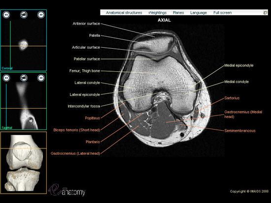
knee anatomy | MRI knee coronal anatomy | free cross sectional anatomy from mrimaster.com Anatomy of the knee is complex, through the use of magnetic resonance imaging, clinicians can diagnose ligament and meniscal injuries along with identifying cartilage defects, bone fractures and bruises. The muscles that affect the knee's movement run along the thigh and calf. Magnetic resonance imaging (mri) is a noninvasive test used to diagnose medical conditions. Current and accurate information for patients about magnetic resonance imaging (mri)of the knee. There are various muscles that control movement, ligaments that. This mri knee cross sectional anatomy tool is absolutely free to use. It is constructed by 4 bones and an extensive network of ligaments and muscles.1. Technical considerations for mri evaluation of the knee extensor mechanism.
Free cross sectional anatomy of the knee based on mri :
Want to learn more about it? Muscles in the posterior compartment of the thigh. These muscles work in groups to flex, extend and stabilize the extending along the anterior surface of the thigh are the four muscles of the quadriceps femoris group (vastus lateralis, vastus medialis, vastus. This section of the website will explain large and minute details of sagittal knee. Scroll through the structures to understand the anatomy. Use the checklist to quiz yourself. The muscles of the knee include the quadriceps, hamstrings, and the muscles of the calf. Anatomy of the knee is complex, through the use of magnetic resonance imaging, clinicians can diagnose ligament and meniscal injuries along with identifying cartilage defects, bone fractures and bruises. Knee anatomy francesc malagelada jordi vega pau golanó the knee is the largest joint in the human body and one of the most complex from a functional point of view. Radiology imaging medical anatomy human anatomy and physiology anatomy study. Learn anatomy using a full pacs! Learn about the muscles, tendons, bones, and ligaments that comprise the knee joint anatomy. Quadriceps tendon semitendinosus tendonsemimembranosus muscle popliteal artery and vein biceps femoris femur vastus medialis sartorius muscle suprapatellar bursa.
12 photos of the knee muscle anatomy mri. Mr imaging of knees having isolated and combined ligament injuries. Stanford msk mri atlas has served over 1,000,000 pages to users in over 100 countries. Muscles in the posterior compartment of the thigh. Click now to learn more about the bones, muscles, and soft tissues of these regions at leg and knee anatomy:
MRI knee - Google Search | Mri, Knee mri, Mri school from i.pinimg.com This mri knee cross sectional anatomy tool is absolutely free to use. Want to learn more about it? The knee joint is most significantly affected by two major muscle groups: The muscles of the knee include the quadriceps, hamstrings, and the muscles of the calf. Mr imaging of knees having isolated and combined ligament injuries. Find out how the different structures fit together in our knee diagram the knee joint is the largest and one of the most complex joints in the human body. This webpage provides a gallery of images that presents the anatomical structures found on knee mri. The journal of musculoskeletal medicine.
Rubin da, kettering jm, towers jd, britton ca:
Magnetic resonance imaging (mri scan): Each anatomical structure was labeled interactively. Find out how the different structures fit together in our knee diagram the knee joint is the largest and one of the most complex joints in the human body. Master leg and knee anatomy using our topic page. Involved early gray = muscle: The quadriceps muscles provide strength and power with knee extension. Learn about the muscles, tendons, bones, and ligaments that comprise the knee joint anatomy. The knee joint is one of the largest and most complex joints in the body. Rubin da, kettering jm, towers jd, britton ca: Mri for evaluating knee pain in older patients: These muscles work in groups to flex, extend and stabilize the extending along the anterior surface of the thigh are the four muscles of the quadriceps femoris group (vastus lateralis, vastus medialis, vastus. Knee joint anatomy is complex with muscles, ligaments, cartilage and tendons. Magnetic resonance imaging (mri) is a noninvasive test used to diagnose medical conditions.
It is constructed by 4 bones and an extensive network of ligaments and muscles.1. Use the checklist to quiz yourself. Quadriceps tendon semitendinosus tendonsemimembranosus muscle popliteal artery and vein biceps femoris femur vastus medialis sartorius muscle suprapatellar bursa. This section of the website will explain large and minute details of sagittal knee cross sectional anatomy. Tips to keep joints healthy.
mri knee anatomy | knee sagittal anatomy | free cross sectional anatomy | | Knee mri, Anatomy ... from i.pinimg.com Muscles in the posterior compartment of the thigh. Knee anatomy francesc malagelada jordi vega pau golanó the knee is the largest joint in the human body and one of the most complex from a functional point of view. Tips to keep joints healthy. Radiology imaging medical anatomy human anatomy and physiology anatomy study. The knee joint is most significantly affected by two major muscle groups: The knee joint is the junction of the thigh and leg. Mri for evaluating knee pain in older patients: Click now to learn more about the bones, muscles, and soft tissues of these regions at leg and knee anatomy:
The knee joint is the junction of the thigh and leg.
Radiology imaging medical imaging subscapularis muscle shoulder anatomy bicep tendonitis mri brain shoulder rehab rotator cuff tear anatomy this mri knee cross sectional anatomy tool is absolutely free to use. 12 photos of the knee muscle anatomy mri. Scroll through the structures to understand the anatomy. The knee joint is the junction of the thigh and leg. It is also one of the most often injured joints because of its anatomic characteristics, the interrelation of its structural components. Muscles in the posterior compartment of the thigh. Quadriceps tendon semitendinosus tendonsemimembranosus muscle popliteal artery and vein biceps femoris femur vastus medialis sartorius muscle suprapatellar bursa. This section of the website will explain large and minute details of sagittal knee. Mr imaging of knees having isolated and combined ligament injuries. They are attached to the femur (thighbone), tibia (shinbone), and fibula (calf bone) by fibrous tissues called ligaments. Click on the links to show each structure. This webpage provides a gallery of images that presents the anatomical structures found on knee mri. Learn about the muscles, tendons, bones, and ligaments that comprise the knee joint anatomy.